IGF-1 LR3 identity is confirmed first by intact mass: about 9,111 Da for the 83-residue analog with three disulfide bonds. Mass tells it apart from native IGF-1 and shows the disulfides formed. It cannot show which cysteines pair. Peptide mapping, run reduced and non-reduced, answers that.
- IGF-1 LR3 has 83 residues, six cysteines forming three disulfides, four methionines and no tryptophan.
- The folded analog comes to about 9,111 Da by average residue masses, about 6 Da below the reduced chain.
- Intact mass separates LR3 from native IGF-1 and from Long IGF-1, but not from misfolded forms with a different disulfide pairing.
- ICH Q6B names peptide mapping under reducing and non-reducing conditions for locating disulfide bridges.
- Each oxidized methionine adds about 16 Da to the observed mass.
Why identity is its own question
How do you tell a protein from its near-twin? IGF-1 LR3 differs from a sibling analog, Long IGF-1, at exactly one position out of 83. Both are white powders. Both dissolve the same way. A purity test would call either one 98 percent pure without blinking, because purity only measures how much of the sample is one main component. It never asks which component that is.
Identity is the separate question: is the molecule in this vial the one on the label? ICH Q6B, the international guideline on specifications for biotechnological products, says an identity test should be highly specific and based on unique aspects of the molecule's structure, and that more than one test may be needed. For a disulfide-bonded protein of this size, that advice applies directly.
This page walks through what a laboratory can check, what each check can and cannot tell apart, and how to read the identity section of a certificate for this analog. The companion page on the IGF-1 LR3 certificate of analysis covers the rest of the document.
- Settle the target: 83 residues, Arg at IGF-1 position 3
- Intact mass near 9,111 Da
- Reduce and remeasure: about 6 Da higher
- Check for plus 16 oxidation peaks
- Peptide map, non-reduced and reduced
- Confirm the N-terminal extension
The target: what the sequence should be
Every identity check compares a measurement against a target, so the target has to be settled first. UniProt entry P05019 gives mature human IGF-1 as 70 residues, starting Gly-Pro-Glu. IGF-1 LR3 replaces that glutamic acid at position 3 with arginine and adds a 13-residue extension in front, Met-Phe-Pro-Ala-Met-Pro-Leu-Ser-Ser-Leu-Phe-Val-Asn. Francis and colleagues described both changes in two 1992 papers, the extension being built from the first 11 residues of methionyl porcine growth hormone.
| Feature | Count or position | Why it matters |
|---|---|---|
| Residues | 83 (13 extension plus 70 IGF-1) | Sets the theoretical mass |
| Arginine substitution | Position 16 in analog numbering, position 3 of the IGF-1 portion | The one residue that separates it from Long IGF-1 |
| Cysteines | Six, paired as 19-61, 31-74 and 60-65 | Three disulfides; pairing must be confirmed separately from mass |
| Methionines | Four (two in the extension, two in the IGF-1 portion) | Oxidation adds 16 Da each and can confuse a mass match |
| Tryptophan and tyrosine | No tryptophan, two tyrosines | Limits UV absorbance at 280 nm |
The disulfide positions come from UniProt's record for IGF-1 (Cys54-Cys96, Cys66-Cys109 and Cys95-Cys100 in precursor numbering), shifted to count from the start of the analog.
What intact mass should IGF-1 LR3 show?
IGF-1 LR3 should show an intact mass near 9,111 Da for the folded form. Mass spectrometry weighs the whole molecule. By our sum of average residue masses, the reduced 83-residue chain comes to about 9,117 Da, and the folded form with three disulfides about 9,111 Da, since each disulfide removes two hydrogens, as PubChem's formula for cystine shows. Q6B lists mass spectrometry among the standard ways to determine molecular weight.
Mass does a lot of work here. It separates the analog cleanly from native IGF-1, which is near 7.6 kDa by the same arithmetic. It shows whether the disulfides have formed, through that six-unit difference. A laboratory can make the point sharper by reducing a portion of the sample, for example with dithiothreitol, and measuring again: the mass should rise by about 6 Da as the bonds open.
It also flags modifications. Each oxidized methionine adds about 16 Da, so a peak 16 or 32 units above the main one is a sign of oxidized material rather than a different sequence.
What can intact mass not tell apart?
Intact mass cannot tell which cysteines pair with which, and it can miss changes that barely move the weight. Swapping the glutamic acid at position 3 of IGF-1 for arginine changes the mass by about 27 Da (arginine's residue mass minus glutamic acid's). A good spectrometer resolves that easily on a 9 kDa protein, so Long IGF-1 and IGF-1 LR3 can be told apart by mass. Other confusions are harder.
- Sequence order. Two chains with the same residues in a different order have the same mass. Intact mass confirms composition, not sequence.
- Disulfide pairing. Six cysteines can form three bonds in fifteen different pairings. Every one of them weighs exactly the same. A misfolded protein with the wrong pairing passes an intact mass check.
- Coincidental matches. Leucine and isoleucine have identical masses, so a swap between them is invisible to intact mass.
That is why ICH Q6B allows that more than one test may be needed. Intact mass is a strong first filter. It is not the whole answer for a folded protein.
How is disulfide pairing confirmed?
Disulfide pairing is confirmed by peptide mapping, which looks inside the molecule. Q6B describes it as selective fragmentation of the product into discrete peptides, using suitable enzymes or chemicals, followed by HPLC or another suitable method. Each fragment is identified, often by mass spectrometry, and the pattern is compared with what the sequence predicts. A substitution anywhere in the chain shows up as a fragment with the wrong mass.
For disulfides the guideline is specific. Where the sequence says cysteines are expected, the number and positions of any free sulfhydryl groups and disulfide bridges should be determined, to the extent possible, and peptide mapping under reducing and non-reducing conditions is one of the methods named. The logic is easy to follow. Under non-reducing conditions, fragments joined by a disulfide stay linked and appear as one larger mass. After reduction they fall apart into their separate pieces. Matching the linked pairs to 19-61, 31-74 and 60-65 confirms the pairing.
Q6B also mentions comparing the terminal amino acid sequence with the sequence expected from the gene. For this analog, confirming that the chain starts with Met-Phe-Pro-Ala is a direct check on the extension. Our guide to LC-MS identity testing covers the instruments in more depth.
Checks a receiving laboratory can run
Not every lab has a mass spectrometer, and a few simpler methods still add something. Q6B lists size exclusion chromatography and SDS-polyacrylamide gel electrophoresis, under reducing and non-reducing conditions, alongside mass spectrometry as ways to determine molecular weight or size.
A gel will not resolve a 27 Da difference, so it cannot separate LR3 from Long IGF-1. What it can show is gross trouble: a band far from the 9 kDa region, a ladder of breakdown products, or higher bands that point to chains linked to each other through stray disulfides. Running the same sample with and without a reducing agent is informative for the same reason. Material held together by bonds between chains changes size when those bonds are broken.
Size exclusion chromatography answers a related question, whether the protein is present as single molecules or as aggregates. Neither method confirms the sequence. Both are cheap ways to catch a problem before a longer experiment depends on the material.
Reading the identity section of a certificate
Most research certificates report identity as a single intact mass. That is a reasonable minimum. When you read one for IGF-1 LR3, check these points.
- The theoretical mass matches the 83-residue analog with three disulfides, near 9,111 Da, not native IGF-1 and not the reduced chain.
- The observed mass sits within the tolerance the method states.
- The spectrum is attached, so you can see whether there are satellite peaks at plus 16 or plus 32.
- The method is named, electrospray or MALDI-TOF, with the ions used to derive the mass.
Peptide mapping or disulfide confirmation on a research certificate is uncommon. If your work depends on correct folding, it is fair to ask whether it was done and, if not, to plan for it in your own laboratory. A certificate that claims identity from HPLC retention time alone has not shown identity at all; see HPLC purity versus identity for why.
Where our certificates stand
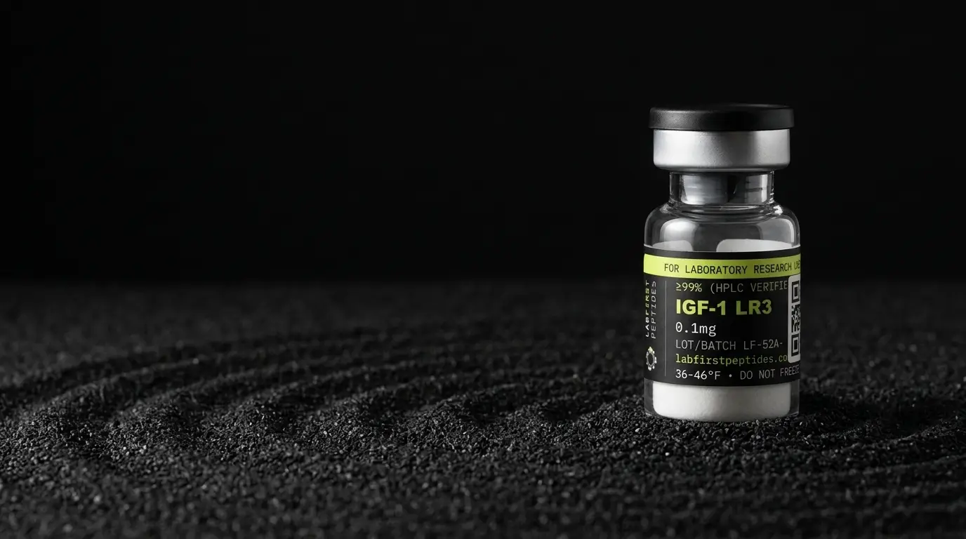
Identity results for our IGF-1 LR3 lots, the 0.1 mg vial (SKU IG01) and the 1 mg vial (SKU IG1), are pending. This page states no result for any of our lots, and a lot's results appear only once the certificate for that lot is published. The IGF-1 LR3 product page and the certificate index will carry them when released. Disulfide bonds and methionines also shape how the material should be kept, which is covered in IGF-1 LR3 storage and stability.
IGF-1 LR3 is not an FDA-approved drug product and has no pharmacopeial monograph, so identity is tested against a supplier-defined specification. It is supplied here as laboratory material only.
FOR LABORATORY AND IN-VITRO RESEARCH USE ONLY. NOT FOR HUMAN OR ANIMAL CONSUMPTION.
What is IGF-1 LR3 studied for?
Published research on IGF-1 LR3 investigates the areas below, which is a different question from what IGF-1 LR3 will do for anyone, a claim about a living system that nothing on this site is sold for.
What it is. Analogue of human insulin-like growth factor 1 with arginine replacing glutamate at position 3 and a thirteen-residue N-terminal extension.
What the research looks at. Used principally as a cell-culture reagent — it is a standard supplement in serum-free and reduced-serum media for industrial and research cell culture — and as a comparator in IGF analogue pharmacology.
How it is thought to work. Binds the IGF-1 receptor. The substitutions markedly reduce affinity for IGF binding proteins, which is the design intent: less sequestration by IGFBPs means more free peptide available to the receptor.
What is not established. No approved product and no human therapeutic data. Reduced IGFBP binding is a property demonstrated in binding assays; that it produces greater biological effect does not follow, and in at least one published comparison it did not.
The full record, including the certificate for the lot in stock, is on the IGF-1 LR3 product page.
Common questions
Can mass spectrometry tell IGF-1 LR3 from Long IGF-1?
How do you confirm the disulfide bonds are paired correctly?
What does a peak 16 Da above the main mass mean?
Is HPLC retention time enough to prove identity?
More documentation guides
Sources
- UniProt P05019, Insulin-like growth factor 1 (human). Mature IGF-1 chain (residues 49 to 118 of the precursor, 70 residues), its sequence, and the three disulfide bonds Cys54-Cys96, Cys66-Cys109 and Cys95-Cys100 in precursor numbering.
- Francis et al., Novel recombinant fusion protein analogues of insulin-like growth factor (IGF)-I, Journal of Molecular Endocrinology, 1992. Describes the Long IGF-I analogues built from the first 11 residues of methionyl porcine growth hormone joined to IGF-I, including Long [Arg3]-IGF-I; PMID 1378742.
- Francis et al., Production and characterization of recombinant IGF-I and potent analogues of IGF-I, Journal of Molecular Endocrinology, 1992. Replacement of glutamic acid at position 3 with arginine or glycine, and reduced binding to IGF-binding proteins as the design rationale; PMID 1311930.
- ICH Q6B, Specifications: Test Procedures and Acceptance Criteria for Biotechnological/Biological Products. Identity tests should rest on unique aspects of molecular structure; where cysteines are expected, free thiols and disulfide bridges should be determined, for example by peptide mapping under reducing and non-reducing conditions or mass spectrometry.
- PubChem compound records for L-cysteine (CID 5862) and L-cystine (CID 67678). Cysteine is C3H7NO2S; cystine, two cysteines joined by a disulfide, is C6H12N2O4S2, two hydrogens fewer than two free cysteines.