Gln-Met-Cys is a linear tripeptide, H-Gln-Met-Cys-OH, formula C13H24N4O5S2, average mass 380.48 Da. Two α-peptide bonds join glutamine, methionine and cysteine. Its analytical behavior is dominated by three reactive groups: a free thiol, a thioether, and an N-terminal glutamine that cyclises to pyroglutamate.
- Gln-Met-Cys is H-Gln-Met-Cys-OH, formula C13H24N4O5S2, average mass 380.48 g/mol and monoisotopic mass 380.1188 Da.
- The peptide has no aromatic residue, so it is detected at 210 to 220 nm and its concentration cannot be determined by absorbance at 280 nm.
- The N-terminal glutamine cyclizes to pyroglutamate with loss of ammonia, a 17 Da shift that runs in aqueous solution without any added reagent.
- The free cysteine thiol forms a symmetric disulfide dimer with a calculated protonated mass of 759.2292, usually the largest single impurity.
- Sequence permutations such as Cys-Met-Gln share the identical exact mass, so identity confirmation requires MS/MS fragment assignment rather than intact mass alone.
- L-cysteine carries the (R) descriptor because sulfur outranks the carboxyl carbon under CIP rules, which is not evidence of a D-residue.
What this guide covers
In plain terms, Gln-Met-Cys is three amino acids linked in a row, like three beads on a string in a fixed order. Swap the order and you get a different molecule. The name tells you which bead comes first and which comes last. This guide covers what the name pins down, how each residue can degrade, how to tell the compound from its isomers and from glutathione, how a laboratory verifies it, and how it should be stored and recorded.
- N-terminal glutamine — cyclises to pyroglutamate, −17 Da
- Glutamine side chain — deamidates to glutamate, +1 Da
- Methionine thioether — oxidises to sulfoxide, +16 Da
- Cysteine thiol — dimerises to disulfide, doubling the observed mass
- Over-oxidised thiol — sulfinic and sulfonic acid, +32 and +48 Da
What does the name Gln-Met-Cys specify?
Gln-Met-Cys is a linear tripeptide written in the standard three-letter code and read from the amino terminus to the carboxy terminus. Glutamine occupies position one, methionine position two, cysteine position three. In one-letter code it is QMC. Unless a supplier states otherwise, that notation carries four silent assumptions: every residue is the L enantiomer, the residues are joined α-carboxyl to α-amino, the N-terminus is a free amine and the C-terminus is a free acid. Spelled out, the material is H-Gln-Met-Cys-OH.
Those are conventions, and conventions get broken quietly. A C-terminal amide would be written -NH2. A side-chain-linked glutamyl residue would need an explicit γ. A catalog entry reading only GMC tripeptide tells you nothing about which of several possible isomers is in the vial, and the difference is not cosmetic.
The systematic name is (2R)-2-{[(2S)-2-{[(2S)-2-amino-4-carbamoylbutanoyl]amino}-4-(methylsulfanyl)butanoyl]amino}-3-sulfanylpropanoic acid. The R descriptor on the cysteine stereocenter is correct and does not indicate D-cysteine. Sulfur outranks the carboxyl carbon under Cahn-Ingold-Prelog priority rules, so L-cysteine is formally (R) while L-glutamine and L-methionine are (S). This trips people up on a regular basis when they read an IUPAC name off a supplier page and conclude the wrong enantiomer was shipped.
| Property | Value |
|---|---|
| Sequence (N to C) | L-Gln - L-Met - L-Cys |
| One-letter code | QMC |
| Molecular formula | C13H24N4O5S2 |
| Average molecular mass | 380.48 g/mol |
| Monoisotopic mass | 380.1188 Da |
| [M+H]+ (calculated) | 381.1261 |
| [M+Na]+ (calculated) | 403.1080 |
| Disulfide-linked dimer, [M+H]+ | 759.2292 |
| Aromatic residues | None; no useful absorbance at 280 nm |
How do its bonds and charge affect column behavior?
Two amide bonds hold the molecule together. Both are planar and both sit overwhelmingly in the trans configuration, since there is no proline to make cis-trans isomerism a practical concern. The backbone is short enough that the peptide has no meaningful secondary structure in solution; conformational questions that dominate the handling of a 39-residue peptide are absent here.
Charge is where the interesting behavior starts. Three groups titrate across the accessible pH range: the α-amino group of glutamine near pH 9, the C-terminal carboxyl near pH 3, and the cysteine thiol somewhere around 8.5 in a peptide context. The glutamine side-chain carboxamide is neutral throughout and does not titrate. Net charge is therefore approximately zero between about pH 4 and pH 8, and the isoelectric point falls near 6.
Two practical consequences follow. Ion-exchange chromatography has a poor handle on this molecule at working pH, so reversed-phase with an ion-pairing additive is the default analytical mode. And the peptide is small and polar, so it elutes early. A conventional C18 run at low organic content may barely retain it. Polar-endcapped or aqueous-compatible C18 phases hold it better, and HILIC is a reasonable orthogonal method when a second confirmation of identity is wanted. Free thiols also interact with stainless steel flow paths, which shows as tailing that improves noticeably on a bio-inert or PEEK-lined system.
How can a Gln-Met-Cys sample degrade?
This sequence packs an unusual density of chemical liability into three residues. Each side chain has a well-documented failure route, and all three are active under ordinary bench conditions.
The N-terminal glutamine is the one people forget. Its free α-amino nitrogen can attack the side-chain amide carbonyl intramolecularly, closing a five-membered lactam and expelling ammonia. The product is an N-terminal pyroglutamyl residue, 17 Da lighter, chromatographically distinct and no longer available for Edman degradation. The reaction runs in aqueous solution at neutral and mildly acidic pH and accelerates with temperature. Material shipped as a solution, or held wet for weeks, will show it.
Methionine oxidizes to the sulfoxide on contact with air, faster in light and faster again in the presence of trace peroxide or metal ions. The cysteine thiol oxidizes to the symmetric disulfide dimer, which is the most common single impurity in any thiol-containing peptide and the reason vials are often filled under inert gas. Over-oxidation continues past the disulfide to sulfinic and sulfonic acids.
| Modification | Site | Δ monoisotopic | Modified [M+H]+ | Typical trigger |
|---|---|---|---|---|
| Pyroglutamate formation | N-terminal Gln | −17.0265 | 364.0996 | Aqueous storage, heat, time |
| Deamidation to Glu | Gln side chain | +0.9840 | 382.1101 | Alkaline pH, heat |
| Methionine sulfoxide | Thioether | +15.9949 | 397.1210 | Air, light, trace peroxide |
| Methionine sulfone | Thioether | +31.9898 | 413.1159 | Strong oxidant |
| Disulfide dimer | Two Cys thiols | 2M − 2.0157 | 759.2292 | Dissolved oxygen, metals, pH above 7 |
| Cysteine sulfinic acid | Thiol | +31.9898 | 413.1159 | Over-oxidation |
| Cysteine sulfonic acid | Thiol | +47.9847 | 429.1108 | Over-oxidation |
| Mixed disulfide with glutathione | Thiol | +305.0682 | 686.1943 | Thiol exchange in a buffer containing GSH |
Two rows in that table share a mass shift. Methionine sulfone and cysteine sulfinic acid both add 31.9898, and intact-mass measurement cannot separate them at any resolution. Localising the oxygens needs MS/MS and inspection of which fragment carries the shift.
How do you tell Gln-Met-Cys from its isomers and from glutathione?
Cys-Met-Gln has the same molecular formula as Gln-Met-Cys, the same average mass and the same exact mass. So does Met-Gln-Cys, and so does any other permutation of the three residues with α-linkages and free termini. High-resolution mass spectrometry confirms elemental composition and says nothing about order. Sequence confirmation requires fragmentation and an examination of the b and y series.
| Ion | Composition | Calculated m/z | Diagnostic value |
|---|---|---|---|
| b2 | Gln-Met | 260.1069 | Confirms Gln precedes Met |
| a2 | b2 − CO | 232.1120 | Supporting confirmation of b2 |
| y2 | Met-Cys | 253.0681 | Confirms Met precedes Cys |
| y1 | Cys | 122.0276 | Places cysteine at the C-terminus |
| Met immonium | — | 104.0534 | Presence of methionine |
| b2 of reverse sequence | Cys-Met | 235.0575 | Absent if the sequence is QMC |
A separate confusion is worth heading off. Glutathione is also a tripeptide, also carries a free cysteine thiol, and is written γ-Glu-Cys-Gly. Its formula is C10H17N3O6S and its monoisotopic mass is 307.0838. The glutamyl residue in glutathione links through its side-chain γ-carboxyl, which is why the γ prefix is mandatory and why the molecule resists ordinary aminopeptidases. Gln-Met-Cys is a different compound with a different linkage pattern, a different residue at position two and an amide rather than a carboxylic acid on the first side chain. Superficial structural resemblance between two small thiol peptides is not a shared biochemistry, and any claim connecting the two needs data attached to it rather than an analogy.
How is Gln-Met-Cys verified, step by step?
Identity and purity for a peptide this size are established by a small, unglamorous set of methods. None of them is exotic and all of them should appear on a competent certificate.
- Run reversed-phase HPLC at 214 nm. Reversed-phase HPLC is the workhorse. A C18 column, a water/acetonitrile gradient with 0.1% trifluoroacetic acid, and detection at 214 nm. The wavelength is not a stylistic choice. Glutamine, methionine and cysteine carry no aromatic ring, so the molecule has no useful chromophore at 280 nm and detection depends on the amide bonds absorbing in the far UV. That also means concentration cannot be determined by A280. Quantitation falls to amino acid analysis, gravimetry corrected for peptide content, or nitrogen determination.
- Run it again after reduction. Run the sample twice: once as prepared, once after treatment with a reducing agent such as TCEP. If a late-eluting peak collapses into the main peak on reduction, that peak was the disulfide dimer, and the unreduced chromatogram was quietly reporting oxidized material as an impurity of unknown identity. This single comparison resolves more purity arguments than any other test.
- Measure free thiol directly. Free thiol content is measured directly with Ellman's reagent, DTNB, which releases the TNB dianion with an absorbance maximum near 412 nm and a molar absorptivity in the region of 14,000 M−1cm−1 at mildly alkaline pH. Comparing measured thiol against theoretical thiol per mole gives an oxidation state figure that HPLC area percent does not.
- Check composition and stereochemistry. Amino acid analysis has one caveat specific to this sequence. Standard 6 M HCl hydrolysis at 110 °C destroys cysteine unless the sample is first oxidized to cysteic acid with performic acid, and it hydrolyzes the glutamine side chain to glutamate. An analysis report showing Glx and no Cys is behaving normally, not signaling a wrong compound. Stereochemistry is checked separately by derivatizing the hydrolysate with Marfey's reagent and looking for D-epimers. In 1H NMR, the methionine S-methyl singlet near 2.1 ppm is the easiest single confirmation that the second residue is present at all.
What impurities does the synthesis predict?
Material of this kind is made by Fmoc solid-phase synthesis, typically on a Wang or 2-chlorotrityl resin preloaded with the cysteine residue. Both the cysteine thiol and the glutamine side-chain amide are normally trityl-protected. The Gln(Trt) protection is not optional in careful practice: an unprotected carboxamide can dehydrate to a nitrile during carbodiimide activation, giving a −18 Da impurity that is easy to miss.
Cleavage from resin is done with trifluoroacetic acid plus scavengers. Triisopropylsilane and water are standard, and a thiol scavenger such as EDT or DODT is added when a free cysteine is present, because liberated trityl cations will otherwise re-alkylate the thiol. The resulting S-trityl adduct sits about 242.11 Da above the parent mass and is a recognizable signature of a badly designed cleavage cocktail.
Cysteine is also the residue most prone to racemization during coupling, particularly under base-heavy activation conditions, which is the reason the chiral check in the previous section exists. The remaining expected impurities are the ordinary ones: deletion sequences missing methionine or glutamine, truncated dipeptides, the pyroglutamate form, and the oxidation products already tabulated.
Reading the certificate against that list is the point. Chromatographic purity is an area percentage at a stated wavelength on a stated method; peptide content is the fraction of the powder in the vial that is peptide rather than counterion and water. For a TFA salt of a small peptide, net peptide content in the range of 70 to 85 percent is unremarkable, and a certificate reporting 99 percent purity while omitting content is describing a chromatogram, not the contents of the container. Water by Karl Fischer, counterion by ion chromatography, and residual solvents by headspace GC against ICH Q3C limits round out what a full document contains. Our quality standard sets out which of these we require per lot.
What to check on a Gln-Met-Cys certificate
- A catalog entry that reads only
GMC tripeptide, with no statement of which isomer is in the vial. - A purity figure with no peptide content beside it. A 99 percent purity result that omits content describes a chromatogram, and the vial holds counterion and water as well.
- An unreduced chromatogram only. Without the TCEP comparison, a disulfide dimer can pass as an impurity of unknown identity.
- Intact mass offered as proof of sequence. Cys-Met-Gln and the other permutations share the same exact mass, so sequence needs MS/MS.
- A peak about 242.11 Da above the parent, the S-trityl adduct left by a poorly designed cleavage.
- Missing water, counterion or residual solvent results.
One thing that looks like a fault and is not: an amino acid analysis showing Glx and no Cys is normal for standard acid hydrolysis of this sequence.
How should Gln-Met-Cys be stored, and what goes in the record?
Hold the lyophilized solid cold, dark and desiccated in a sealed vial, ideally with an inert headspace given the free thiol. Let a cold vial equilibrate to room temperature before the seal is broken; condensation onto a hygroscopic cake is the most common avoidable handling error, and it is invisible afterwards. The general reasoning is set out in the guide on storage and stability of lyophilized peptides.
In solution, the thiol governs everything. Dissolved oxygen and trace transition metals drive disulfide formation, and the rate rises sharply once the pH climbs past the thiol pKa and thiolate becomes the dominant species. That chemistry is the reason the solid stays sealed and dry.
The record itself is short. Write down the source lot, the date received, where the vial is stored and the initials of whoever checked it. Note the average mass of 380.48 g/mol beside it, since every molar figure depends on that number.
What is the regulatory position of Gln-Met-Cys?
Gln-Met-Cys is a research chemical. As of the verification date below, no FDA-approved drug product lists this tripeptide as an active ingredient, and no pharmacopeial monograph covering the assembled tripeptide could be confirmed. Monographs exist in USP-NF for the free amino acids methionine and cysteine hydrochloride, which govern those raw materials and say nothing about a synthesised peptide made from them.
Supply is therefore governed by the general framework for research chemicals: accurate labeling, a lot-specific certificate of analysis, and no representation of suitability for use in humans or animals. Absence of an approval is not an absence of regulation, and it is certainly not evidence of safety.
Status verified August 26, 2026.
FOR LABORATORY AND IN-VITRO RESEARCH USE ONLY. NOT FOR HUMAN OR ANIMAL CONSUMPTION. NOT FOR PERSONAL, MEDICAL, DIAGNOSTIC, THERAPEUTIC, OR RECREATIONAL USE.
Bottom line
Gln-Met-Cys is H-Gln-Met-Cys-OH, C13H24N4O5S2, 380.48 g/mol. Three groups make it fragile: the N-terminal glutamine cyclizes to pyroglutamate, the methionine oxidizes, and the cysteine thiol forms a disulfide dimer at m/z 759.2292. Confirm it with HPLC at 214 nm run with and without reduction, a free-thiol assay, and MS/MS for sequence, and read purity beside net peptide content. It is a research chemical, and no approved medicine contains it.
What is Glutathione studied for?
Published research on Glutathione investigates the areas below, which is a different question from what Glutathione will do for anyone, a claim about a living system that nothing on this site is sold for.
What it is. A tripeptide, gamma-glutamyl-cysteinyl-glycine — note the unusual gamma peptide bond at the glutamate, which is not a standard alpha linkage.
What the research looks at. Oxidative stress, redox signalling, detoxification, and the regulation of cell proliferation, apoptosis, immune function and fibrogenesis. An enormous biochemical literature.
How it is thought to work. The most abundant non-protein thiol in mammalian tissue. Functions as the principal intracellular redox buffer, a determinant of redox signalling, and a substrate for detoxification of xenobiotics via glutathione S-transferases. It is synthesised inside cells in two ATP-dependent steps, not taken up intact.
What is not established. That supplying glutathione from outside a cell raises glutathione inside it. Synthesis is intracellular and rate-limited by cysteine availability and glutamate cysteine ligase activity, neither of which is addressed by adding the finished tripeptide.
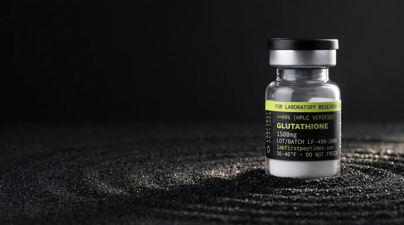
The full record, including the certificate for the lot in stock, is on the Glutathione product page.
Common questions
Is Gln-Met-Cys the same thing as glutathione?
Why does the certificate report purity at 214 nm rather than 280 nm?
What is the peak 16 Da above the expected mass?
A peak appears near m/z 759. What is it?
The IUPAC name says (2R) at the cysteine. Is that D-cysteine?
What does net peptide content mean and why is it lower than the purity figure?
More science guides
Sources
- IUPAC-IUB Joint Commission on Biochemical Nomenclature, recommendations on nomenclature and symbolism for amino acids and peptides. Establishes the three-letter and one-letter code conventions, the N-to-C reading order, and the requirement to mark non-α linkages explicitly, as in γ-Glu.
- Ellman, Archives of Biochemistry and Biophysics, 1959. Original description of DTNB for the quantitation of tissue sulfhydryl groups; underpins the free-thiol assay and the 412 nm TNB absorbance measurement.
- PubChem (NIH/NLM) records for L-glutamine, L-methionine and L-cysteine. Source of the residue formulas, stereodescriptors and monoisotopic masses used to derive the tripeptide formula and mass values in this article.
- USP-NF general chapter on chromatography and ICH Q3C guideline for residual solvents. Define system suitability expectations for HPLC purity determination and the residual solvent limits a certificate of analysis is measured against.