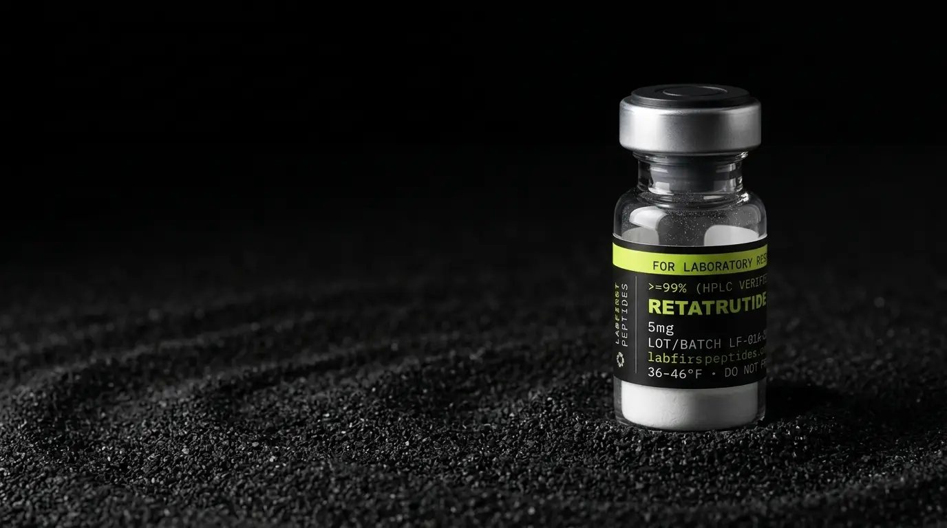
Retatrutide works by binding and switching on three related receptors: the GIP, GLP-1 and glucagon receptors. Each is a class B GPCR that signals by raising cAMP inside the cell. In lab cell assays it is more potent than native GIP at the GIP receptor and about three times less potent than the native hormones at the other two.
- Retatrutide (LY3437943) is a triple agonist peptide at the glucagon, GIP and GLP-1 receptors, per Coskun et al. 2022 in Cell Metabolism.
- PubChem CID 171390338 lists retatrutide's molecular formula as C221H342N46O68, with a molecular weight of about 4,731 g/mol.
- Retatrutide is built on the GIP backbone, with Aib at positions 2 and 20, alpha-methyl leucine at 13, and a C20 fatty diacid on lysine 17.
- A review of glucagon pharmacology reports retatrutide as about 9 times more potent than native GIP and about 3 times less potent than native GLP-1 and glucagon in vitro.
- The glucagon receptor is a 477 amino acid class B GPCR coupled mainly to G-alpha-s, which activates adenylyl cyclase to make cAMP.
- Fatty acid chains on lipidated peptides bind serum albumin through hydrophobic contacts with specific albumin pockets.
What this guide covers
Picture a building with three different doorbells, each wired to the same buzzer inside. Most keys fit one bell. Retatrutide was designed to press all three. The bells are receptors on the surface of a cell, and the buzzer is a small messenger molecule called cAMP.
In the language of the discovery paper, retatrutide (development code LY3437943) is a triple agonist peptide at the glucagon receptor (GCGR), the glucose-dependent insulinotropic polypeptide receptor (GIPR) and the glucagon-like peptide-1 receptor (GLP-1R). An agonist is a molecule that binds a receptor and activates it, rather than just sitting in the way. Retatrutide does this at three receptors with one chain of amino acids.
This page stays at the level where that mechanism has been measured directly: receptors in cultured cells, signaling molecules, and the chemistry that keeps the peptide intact. It runs in this order: what the molecule is made of, which receptors it targets, how the signal reaches cAMP, how potent it is at each receptor, how laboratories measure that, and what the fatty chain on the side does. It closes with a checklist for reading any potency figure, the mistakes people make with mechanism claims, and a bottom line.
- Peptide binds GIPR, GLP-1R or GCGR
- Receptor changes shape inside the membrane
- G-alpha-s activates adenylyl cyclase
- ATP is converted to cAMP
- cAMP is measured and EC50 is read
What is retatrutide made of?
Retatrutide is a modified peptide built on the backbone of GIP, the hormone for one of its three receptors. PubChem lists it under CID 171390338 with the molecular formula C221H342N46O68 and a molecular weight of about 4,731 g/mol. That is roughly ten times the size of a small drug molecule and still small for a protein.
A 2026 review of peptide drugs that act on GPCRs lists the changes made to that GIP backbone:
- Aib (alpha-aminoisobutyric acid) in place of alanine at position 2
- alpha-methyl leucine at position 13
- Aib in place of glutamine at position 20
- a C20 fatty diacid attached to the lysine at position 17 through an AEEA and gamma-glutamic acid linker
- a C-terminal tail borrowed from exendin-4
Each change has a job. The unusual residues near the front of the chain help it resist enzymes that clip peptides, and the fatty chain on lysine 17 is the albumin anchor discussed below. The tail and the substitutions together tune how well the peptide fits each of the three receptors.
Our retatrutide identity guide covers how the formula and mass above are used to confirm the molecule in a vial.
Which receptors does retatrutide target?
Retatrutide targets the GIP, GLP-1 and glucagon receptors, which all belong to class B of the G protein-coupled receptor (GPCR) superfamily, sometimes called the secretin family. The 2026 GPCR review describes class B as receptors for 15 different peptide hormones, with large extracellular domains and large binding sites. Those big binding sites suit peptide ligands, which are much larger than typical small-molecule drugs.
The three receptors are close relatives, and their natural hormones are related peptides too. That family resemblance is what makes a single peptide able to fit more than one of them. A sequence that starts from GIP and borrows features recognized by the GLP-1 and glucagon receptors can, if tuned well, be read as a match by all three.
| Receptor | Native hormone | Receptor class |
|---|---|---|
| GIPR | Glucose-dependent insulinotropic polypeptide (GIP) | Class B GPCR |
| GLP-1R | Glucagon-like peptide-1 | Class B GPCR |
| GCGR | Glucagon | Class B GPCR (secretin family) |
A GPCR crosses the cell membrane seven times. The part outside the cell catches the hormone, and the part inside the cell changes shape when the hormone is bound. That shape change is the signal that passes through the membrane.
How does the signal get from the receptor to cAMP?
Once retatrutide binds, the receptor passes the message to a G protein on the inside of the membrane. An IUPHAR review of glucagon pharmacology describes the glucagon receptor, a 477 amino acid GPCR, as primarily coupled to the G-alpha-s protein, which activates adenylyl cyclase. Adenylyl cyclase is an enzyme that converts ATP into cyclic AMP, or cAMP.
cAMP is a second messenger. The first messenger is the hormone outside the cell; cAMP carries the signal onward inside it. Because cAMP rises when these receptors are switched on, it is the readout laboratories most often use to measure how strongly a peptide activates them.
That shared pathway is a practical convenience. A laboratory can test retatrutide at each of the three receptors with the same kind of assay and the same readout, then compare the numbers side by side. The receptors differ. The downstream messenger being measured is the same.
How potent is retatrutide at each receptor?
The discovery paper by Coskun and colleagues, published in Cell Metabolism in 2022, summarizes the in vitro result in one line: LY3437943 shows balanced GCGR and GLP-1R activity but more GIPR activity. Put simply, it is about equally active at the glucagon and GLP-1 receptors, and stronger at the GIP receptor.
The IUPHAR review of glucagon pharmacology puts that in terms of the native hormones. It describes retatrutide as 9 times more potent than endogenous GIP, 3 times less potent than endogenous GLP-1, and 3 times less potent than endogenous glucagon.
| Receptor | Compared with | Reported relative potency |
|---|---|---|
| GIPR | Native GIP | About 9 times more potent |
| GLP-1R | Native GLP-1 | About 3 times less potent |
| GCGR | Native glucagon | About 3 times less potent |
Potency here means how little peptide is needed to get a given response in the assay, usually expressed as an EC50: the concentration that gives half the maximum response. A lower EC50 means higher potency. These are comparisons inside one assay system, so they describe the molecule's receptor profile and nothing beyond the culture dish.
How do researchers measure receptor activity in cells?
Researchers measure it with a cAMP accumulation assay. Cells are engineered to express one human receptor each, then exposed to a range of peptide concentrations, and the cAMP each well produces is measured.
The same Lilly group described this setup for its earlier dual agonist, tirzepatide. In that 2018 paper, HEK293 cells expressing either human GIPR or GLP-1R were used for whole-cell cAMP accumulation assays. Plotting cAMP against concentration gives an S-shaped curve, and the EC50 is read from its midpoint.
One detail in that method matters for any fatty-acid peptide. The tirzepatide paper states that its binding and cAMP assays in HEK293 cells were run in the absence of albumin, to allow direct comparison to the native peptides without the confounding influence of albumin binding. A lipidated peptide that grabs albumin in the assay buffer would look weaker than it is, because less of it is free to reach the receptor.
So when a potency figure is quoted for a lipidated peptide, the assay conditions are part of the number. A figure measured with albumin present and one measured without it cannot be compared directly, and a careful paper says which it used.
How does the C20 fatty diacid bind albumin?
The fatty chain on lysine 17 binds serum albumin, the most abundant protein in blood plasma, by fitting into its fatty acid pockets. A 2026 review of artificial lipidation in the FEBS Journal explains that the interaction between lipidated peptides and albumin is dominated by hydrophobic contacts between the fatty acid chain and specific binding pockets on albumin. Albumin naturally carries fatty acids, so a fatty chain on a peptide fits pockets that already exist.
The same review lays out why that slows clearance. The albumin-peptide complex is much larger than the peptide alone, which reduces filtering by the kidney. Albumin is also rescued from breakdown inside cells by the FcRn receptor, which returns it to circulation, and a bound peptide rides along. Chain length matters too: longer fatty acid chains create stronger albumin interactions. The GPCR review adds that lipidation also gives steric protection against the enzyme DPP-4, which otherwise cuts this class of peptide near its front end.
Binding to albumin is reversible. Only the free fraction reaches a receptor at any moment, which is why cell assays are run without albumin when the goal is to compare the receptor activity of the peptide itself.
Chemically, that leaves retatrutide with two working parts: a peptide chain that does the receptor binding, and a fatty diacid that does the albumin binding. Our retatrutide storage and stability guide covers how the sealed lyophilized material is kept intact before laboratory work.
How to read a retatrutide potency figure: a checklist
Potency numbers get quoted without their context all the time. Run any figure you meet through these six questions before you repeat it.
- Which receptor? Retatrutide has three, and the numbers differ. A figure with no receptor named is a figure with no meaning.
- Compared with what? The review figures above are relative to each native hormone (GIP, GLP-1, glucagon). A comparison against another synthetic peptide is a different number.
- What was the readout? For these receptors it is usually cAMP accumulation, with potency reported as an EC50. Check that the paper says so.
- Was albumin in the buffer? A lipidated peptide looks weaker with albumin present. The tirzepatide method ran without it for exactly that reason.
- Which cells? The published method uses HEK293 cells engineered to express one human receptor each. A different cell line or a mixed population is a different assay.
- Is it in vitro? Everything on this page is. A number from a culture dish describes the receptor profile of the molecule and nothing past that.
Common mistakes when reading mechanism claims
- Treating retatrutide as a GLP-1 peptide only. GLP-1R is one of three targets, and in vitro it is the GIP receptor where the molecule is most active.
- Comparing EC50 values from different assay conditions. With albumin and without albumin are not the same experiment for a peptide carrying a C20 fatty diacid.
- Confusing the two working parts. The peptide chain binds the receptors. The fatty diacid binds albumin. A claim that mixes them up has not read the structure.
- Reading relative potency as a statement about anything outside the dish. The 9-times and 3-times figures are receptor assay results, as the review reports them, and nothing more.
- Taking a nickname for a mechanism. "GLP-3" describes a count of targets, not how the molecule works. Our GLP-3 and retatrutide page covers where that name came from.
- Forgetting the receptors share a pathway. All three signal through G-alpha-s, adenylyl cyclase and cAMP, which is why one assay type can test all three, and why a result at one receptor does not tell you the result at another.
Bottom line
Retatrutide is one 39-residue peptide on a GIP backbone that activates three class B receptors, GIPR, GLP-1R and GCGR, each of which raises cAMP through G-alpha-s and adenylyl cyclase. In cell assays it is strongest at the GIP receptor and roughly balanced at the other two. The C20 fatty diacid on lysine 17 is for albumin binding, not receptor binding, and it is why potency assays are run without albumin. This page is about mechanism only; the identity guide linked above explains the mass and chromatography evidence a certificate should carry, and the retatrutide product page lists the sizes we carry. Lot results appear only once the certificate for that lot is published.
FOR LABORATORY AND IN-VITRO RESEARCH USE ONLY. NOT FOR HUMAN OR ANIMAL CONSUMPTION.
What is Retatrutide studied for?
Published research on Retatrutide investigates the areas below, which is a different question from what Retatrutide will do for anyone, a claim about a living system that nothing on this site is sold for.
What it is. An experimental peptide that works on three incretin receptors at once.
What the research looks at. It shows up in clinical-stage metabolic research under the developer code LY3437943. The published work is about how it behaves at those receptors, how the effect changes with the amount given, and the measurements the sponsor registered its trials to take.
How it is thought to work. It switches on three receptors at the same time: GIP, GLP-1 and glucagon. Everything before it in this family worked on one or two. Hitting all three is why researchers treat it as its own kind of compound rather than a stronger version of what already existed.
What is not established. No regulator has approved it for anything, anywhere. It is still experimental, and nobody has made a finding about whether it is safe, whether it works, or whether it is fit for any purpose outside a registered trial.
The full record, including the certificate for the lot in stock, is on the Retatrutide product page.
Common questions
What is the retatrutide mechanism of action?
Which retatrutide receptor does it bind most strongly?
What does triple agonist mechanism mean?
Why do cell assays for retatrutide leave out albumin?
What is the C20 fatty diacid on retatrutide for?
Published certificates for Retatrutide
Every figure below is read from a report the laboratory issued for that lot; each page carries the PDF and the lab's own verification link.
More science guides
Sources
- Coskun T, et al. LY3437943, a novel triple glucagon, GIP, and GLP-1 receptor agonist: from discovery to clinical proof of concept. Cell Metabolism 34(9):1234-1247 (2022), doi 10.1016/j.cmet.2022.07.013, Europe PMC record. Defines LY3437943 as a triple agonist at GCGR, GIPR and GLP-1R; in vitro, balanced GCGR and GLP-1R activity with more GIPR activity.
- PubChem PUG REST: CID 171390338 molecular formula and weight. Formula C221H342N46O68 and molecular weight 4731 for the retatrutide record.
- IUPHAR review: From foe to friend: Repurposing glucagon to treat obesity and type 2 diabetes. Pharmacological Research (2026). Glucagon receptor as a 477 amino acid secretin-family GPCR coupled to G-alpha-s and adenylyl cyclase; retatrutide relative potency at each receptor.
- Peptide-based therapeutics targeting GPCRs: recent applications in the treatment of metabolic disorders. Frontiers in Pharmacology (2026). Retatrutide sequence modifications on the GIP backbone; class B GPCR features; lipidation and DPP-4 protection.
- Artificial lipidation of proteins and peptides. FEBS Journal (2026). Hydrophobic binding of fatty acid chains to albumin pockets, FcRn recycling, reduced renal clearance, effect of chain length.
- Coskun T, et al. LY3298176, a novel dual GIP and GLP-1 receptor agonist. Molecular Metabolism (2018). Methods: whole-cell cAMP accumulation assays in HEK293 cells expressing human GIPR or GLP-1R, run without albumin.