Dulaglutide identity is confirmed with protein methods, not the quick checks used for small peptides. In plain terms, it is too big for one mass reading to prove. It is a two-chain protein of about 63 kDa, so a lab combines whole and reduced mass, a peptide map of the sequence, and checks of disulfide bonds and size.
- Dulaglutide is a two-chain Fc-fusion protein of approximately 63 kDa, per the FDA label description.
- Dulaglutide identity is confirmed with protein methods: intact and reduced mass, peptide mapping, and size profiles.
- ICH Q6B says identity tests should be highly specific and that more than one may be needed.
- Q6B describes peptide mapping as frequently used to confirm structure for lot release.
- Q6B notes that amino acid composition is less definitive for large proteins than for peptides.
Why the usual peptide check falls short
How do you prove a 63,000-dalton protein is the one on the label? You cannot do it the way you would for a ten-residue peptide, where one mass reading and a sequence you can add up by hand settle most of the question.
For a short synthetic peptide, a mass within tolerance of the theoretical value rules out most wrong answers, because few plausible errors land on the same number. For a protein this size, many small changes shift the mass by amounts that are hard to see against the whole, and the sequence has hundreds of positions where something could differ.
So dulaglutide identity rests on several tests read together. ICH Q6B, the guideline on specifications for biotechnological products, says identity tests should be highly specific, based on unique aspects of the molecular structure, and that more than one test may be needed. That sentence is the plan for this page.
- Intact mass against the theoretical value
- Reduced mass: one chain, about half
- Peptide map with fragment assignments
- Disulfide and chain structure checks
- SEC main peak at the expected size
What is dulaglutide, structurally?
The FDA label for the approved product describes dulaglutide as a fusion protein of two identical chains linked by disulfide bonds. Each chain begins with a GLP-1 analog sequence, which is joined by a small peptide linker to the Fc portion of a modified human immunoglobulin G4 heavy chain.
The label gives the GLP-1 part as 90 percent homologous to native human GLP-1 (7-37), modified near the site that interacts with the enzyme DPP-4. It states that the molecule is made in Chinese hamster ovary cell culture and that its overall molecular weight is approximately 63 kilodaltons.
Every identity method below is testing one of those statements: the size, the two-chain structure, the disulfide links, or the sequence of the GLP-1 analog, linker and Fc.
Intact and reduced mass
Mass spectrometry on the whole protein is the first check. An electrospray instrument produces a series of ions carrying different numbers of charges, and software converts that series into a single neutral mass. For a protein this large the result is compared against the theoretical mass of the claimed structure, within the instrument's stated tolerance.
Reducing the sample adds a second reading. A reducing agent breaks disulfide bonds, including the ones that hold the two chains together, so the reduced spectrum should show one chain at roughly half the intact mass. Q6B lists mass spectrometry and SDS-PAGE under reducing and non-reducing conditions as ways to determine molecular weight.
Read the two together. An intact mass near the expected value with a reduced mass near half of it supports a two-chain molecule of the right size. Either number alone leaves more open. The certificate should say which condition each figure comes from.
Peptide mapping carries the sequence
A peptide map is the method that reads sequence on a protein this size. Q6B describes it as fragmenting the product into discrete peptides with suitable enzymes or chemicals, analyzing the fragments by HPLC or another method, and identifying them by amino acid composition, N-terminal sequencing or mass spectrometry.
The guideline adds that peptide mapping with a validated procedure is frequently used to confirm the desired structure for lot release. In practice, a map shows whether the fragments expected from the GLP-1 analog, the linker and the Fc region are present at the expected masses, and it can place a modification at a specific residue.
A certificate should name the enzyme, show the chromatogram, and state how fragments were assigned. Sequence coverage, the share of the chain accounted for by identified fragments, is the figure that tells you how much of the molecule the map examined.
Disulfide bonds and size
Q6B says that where cysteines are expected from the gene sequence, the number and positions of free sulfhydryl groups and disulfide bridges should be determined, to the extent possible, and that peptide mapping under reducing and non-reducing conditions and mass spectrometry are useful here. For dulaglutide, the interchain disulfide bonds are what make it a two-chain molecule, so this is part of identity, not a side note.
Size-exclusion chromatography and SDS-PAGE add a size profile. Q6B names both, along with mass spectrometry, as ways to determine molecular weight or size. On SEC the intact protein should be the main peak. Q6B defines aggregates as dimers and higher multiples of the product, and they elute before it. The main peak position confirms size; the other peaks belong on the purity side of the report.
What don't these methods settle?
A matching intact mass and a clean map do not measure how much protein is in the vial, and they do not measure purity. Q6B treats purity separately and says it is usually estimated by a combination of methods, because absolute purity of a biological product is hard to determine.
Amino acid composition is also weaker here than for a small peptide. Q6B notes that composition analysis gives useful structural information for peptides and small proteins but is generally less definitive for large proteins. A dulaglutide certificate that offers amino acid analysis as its identity test has chosen a method the guideline itself calls limited for molecules like this.
The dulaglutide certificate guide walks through the purity and content fields in order.
Reading the identity section of a certificate
| Method | What it confirms | What to look for on the report |
|---|---|---|
| Intact mass (MS) | Overall size of the two-chain protein | Observed and theoretical mass side by side |
| Reduced mass (MS) | Single-chain mass after disulfides are broken | Condition stated as reduced |
| Peptide map | Sequence, and location of modifications | Enzyme, chromatogram, fragment assignments, coverage |
| Non-reduced and reduced SDS-PAGE | Chain structure and size | Both lanes shown |
| SEC | Size of the main species | Main peak with high and low molecular weight species reported |
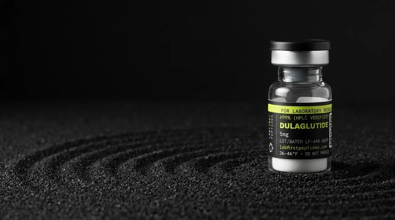
Our certificates are pending, and a lot's results are shown only once the certificate for that lot is published on the certificate index. Nothing on this page reports a result for any of our lots. The dulaglutide product page lists the DL5 (5 mg) and DL10 (10 mg) vials and links a certificate once one exists.
The GLP-1 end of the chain
The part of the molecule that gives dulaglutide its name sits at the N-terminus of each chain. The label says this GLP-1 analog is 90 percent homologous to native GLP-1 (7-37), with changes placed near the site the enzyme DPP-4 interacts with. Those few changed residues are what separate the analog from the native sequence.
An intact mass cannot show them clearly. Swapping one residue for another may move the total by a few tens of daltons against a mass above 60,000, which is similar in size to the shifts that oxidation or deamidation elsewhere on the protein produce. The peptide map is where the difference shows, because the N-terminal fragment is small enough that a single substitution moves its mass by a clearly measurable amount.
So when you read a map, find the N-terminal fragment first. Its observed mass, and whether it matches the modified GLP-1 analog rather than the native sequence, is the most direct evidence that the lot carries the right front end.
Checks a receiving laboratory can run
Not every laboratory has a peptide mapping workflow, and it does not need one to catch the gross errors. A non-reducing and reducing SDS-PAGE pair is inexpensive and shows whether the material runs as a two-chain protein that splits into single chains. An analytical SEC run shows whether the main species elutes where a protein of this size should.
Where results will be published, an intact mass on an in-house or contract instrument is reasonable incoming practice. Record the lot, the date, the method and the observed values in the same notebook entry as the certificate reference, so the chain from vial to data can be followed later.
Regulatory position
Dulaglutide is the active ingredient of an FDA-approved drug product first approved in the United States in 2014. Research material is not that product and is not tested under its approval. Confirming identity by the methods above establishes chemistry, nothing more.
For storage of the sealed vial and of solutions prepared for assays, see dulaglutide storage and stability.
Status verified September 29, 2026.
FOR LABORATORY AND IN-VITRO RESEARCH USE ONLY. NOT FOR HUMAN OR ANIMAL CONSUMPTION.
What is Dulaglutide studied for?
Published research on Dulaglutide investigates the areas below, which is a different question from what Dulaglutide will do for anyone, a claim about a living system that nothing on this site is sold for.
What it is. Recombinant fusion protein: two GLP-1 analogue peptides linked to a modified human IgG4 Fc fragment. Not a peptide in the sense the rest of this catalog uses the word.
What the research looks at. An approved medicine, marketed as Trulicity, with an extensive clinical literature. Also used as a GLP-1 receptor agonist reference compound in preclinical research.
How it is thought to work. Agonist at the GLP-1 receptor. The Fc fusion is the long-acting strategy — a large protein is cleared slowly by the kidney and is recycled through the neonatal Fc receptor, which extends circulating time by a completely different route from the albumin-binding acylation used by semaglutide.
What is not established. That a research vial under this name contains the same molecule as the licensed product. A recombinant fusion protein is manufactured in cell culture rather than synthesised, and identity and purity for such a product mean something different from what they mean for a synthetic peptide.
The full record, including the certificate for the lot in stock, is on the Dulaglutide product page.
Common questions
Can one mass reading confirm dulaglutide identity?
Why does a reduced mass come out at about half?
Is amino acid analysis a good identity test here?
Does SEC confirm identity or purity?
More documentation guides
Sources
- TRULICITY (dulaglutide) prescribing information, section 11 Description (DailyMed, National Library of Medicine). Two identical disulfide-linked chains, GLP-1 analog linked to a modified IgG4 Fc, 90 percent homology to GLP-1 (7-37), CHO expression, approximately 63 kDa.
- ICH Q6B, Specifications: Test Procedures and Acceptance Criteria for Biotechnological/Biological Products. Identity tests, peptide mapping, disulfide determination, molecular weight by SEC, SDS-PAGE and MS, aggregates, limits of amino acid composition for large proteins.