Disulfide bonds in peptides are covalent sulfur-sulfur links between two cysteine residues. Each cysteine gives up the hydrogen on its thiol, so every bond lowers the peptide's mass by about 2.016 Da. The link can sit within one chain or tie two chains together, and a lab breaks it with a reducing agent such as DTT or TCEP.
- LabFirst Peptides certificates report identity by LC-MS, purity by RP-HPLC on a C18 column (most at 214 nm) and net content as a separate line, each by lot number, with a link to the issuing laboratory's site.
- A disulfide bond in a peptide forms when two cysteine thiols are oxidized to cystine, each giving up one hydrogen atom.
- Each disulfide bond removes about 2.016 Da: PubChem lists two L-cysteines at 242.0395 Da combined and cystine at 240.0238 Da.
- Oxytocin's nine-residue sequence Cys-Tyr-Ile-Gln-Asn-Cys-Pro-Leu-Gly carries one disulfide between Cys1 and Cys6, per UniProt P01178.
- Human insulin carries three disulfides, two interchain (B7 to A7 and B19 to A20) and one within the A chain (A6 to A11), per UniProt P01308.
- Thiol-disulfide exchange is highly favored at alkaline pH, so disulfide mapping digests are run at neutral or slightly acidic pH to avoid scrambling the pairings.
What this guide covers
Cysteine is the one standard amino acid with a sulfur-hydrogen group on its side chain. Put two cysteines near each other in a peptide, take one hydrogen off each, and the two sulfurs bond to one another. The chain now has a loop in it, or two chains are stapled together, and the whole thing weighs a little less than it did.
That is a disulfide bond. This guide covers how it forms, the exact mass change it causes, the difference between a bridge inside one chain and a bridge between two, how a laboratory opens the bond on purpose, and how it works out which cysteine is paired with which. Two sourced examples run through it: oxytocin, with one bridge, and insulin, with three.
If you want the broader family of ring-shaped peptides, our guide to cyclic peptides treats the disulfide ring as one of three ways to close a loop. This page stays on the bond itself.
- Two cysteines: each side chain carries a sulfur-hydrogen thiol
- Oxidation: both hydrogens leave and the sulfurs bond
- Mass check: the peptide reads 2.016 Da lighter by LC-MS
- Reduction: DTT or TCEP returns the hydrogens and opens the bond
- Mapping: a non-reduced digest shows which cysteines were joined
What is a disulfide bond?
A disulfide bond is a covalent bond between two sulfur atoms. In a peptide those sulfurs come from the side chains of two cysteine residues, and the paired unit is called cystine. The free amino acid makes the arithmetic plain. PubChem lists L-cysteine as C3H7NO2S with a monoisotopic mass of 121.0197 Da, and cystine as C6H12N2O4S2 at 240.0238 Da. Two cysteines add up to 242.0395 Da. Cystine is 2.0157 Da lighter: the two hydrogen atoms that left, by monoisotopic mass. At the average atomic mass PubChem's periodic table gives hydrogen, 1.0080 Da, the same two atoms come to 2.016 Da.
In plain terms, forming the bond is an oxidation. The LibreTexts organic chemistry text puts it in one sentence: the free dithiol form is the reduced state and the disulfide form is the oxidized state. Breaking the bond is the reverse, a reduction that hands each sulfur its hydrogen back.
The bond is also reversible in a way the peptide backbone is not. A thiol can attack an existing disulfide, take one sulfur for itself and release the other as a new thiol. LibreTexts describes this thiol-disulfide exchange as two SN2-like displacements in a row, with sulfur atoms acting as nucleophile, electrophile and leaving group. That exchange is what lets a cell's glutathione keep intracellular proteins in the free-thiol state, and it is also what causes trouble in the lab, as the mapping section explains.
How much mass does a disulfide bond remove?
Each disulfide bond removes two hydrogen atoms and nothing else, so the peptide loses about 2.016 Da per bond. Nothing is added. The sulfurs were already in the sequence; they just bonded to each other instead of to hydrogen.
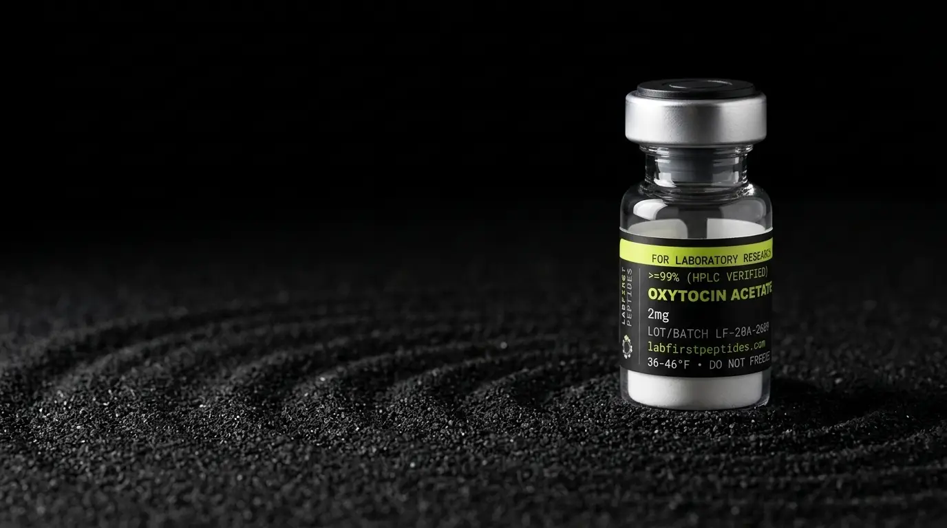
Oxytocin shows it on a real molecule. UniProt entry P01178 gives the nine-residue peptide as Cys-Tyr-Ile-Gln-Asn-Cys-Pro-Leu-Gly with a disulfide between the first and sixth residues and an amide on the C-terminal glycine. PubChem lists the bridged peptide as C43H66N12O12S2 with a monoisotopic mass of 1006.4365 Da. Open the bridge, and the formula becomes C43H68N12O12S2 at about 1008.452 Da. That is our arithmetic from the two records, and the gap is the 2.016 Da of the bond.
| Species | Formula | Monoisotopic mass (Da) |
|---|---|---|
| L-cysteine, one residue | C3H7NO2S | 121.0197 |
| Two cysteines, added | 2 x C3H7NO2S | 242.0395 |
| Cystine, the bonded pair | C6H12N2O4S2 | 240.0238 |
| Difference per bond | H2 | 2.0157 |
| Oxytocin, bridged | C43H66N12O12S2 | 1006.4365 |
| Oxytocin, bridge opened (our arithmetic) | C43H68N12O12S2 | 1008.452 |
Two daltons is small against a thousand, so this is a measurement for a mass spectrometer, not a balance. On a certificate the identity check by LC-MS is where it shows. If the observed mass matches the bridged formula, the bond is closed. If it lands 2 Da high, the cysteines are still free. Our guide to how oxytocin acetate identity is confirmed walks through that exact comparison on a certificate.
Intrachain and interchain disulfides
A disulfide can join two cysteines on the same chain, which folds the chain back on itself into a loop, or it can join cysteines on two separate chains. The Wheaton College biochemistry text on LibreTexts calls these intrachain and interchain bonds, and insulin has both.
UniProt entry P01308 lays out the human insulin precursor as 110 residues: a signal peptide at 1 to 24, the B chain at 25 to 54, a C-peptide at 57 to 87 that is cut away, and the A chain at 90 to 110. The entry records three disulfides in precursor numbering: 31 to 96 and 43 to 109, both marked interchain, and 95 to 100 within the A chain. Renumbering each chain from its own first residue gives B7 to A7, B19 to A20, and A6 to A11. That renumbering is our arithmetic from the chain boundaries in the entry.
Those three bonds are why insulin stays one molecule after the C-peptide is removed. Two chains of 30 and 21 residues are held together by two interchain bridges, and the A chain carries its own short loop. Three bonds also means the mature protein sits about 6.05 Da below the sum of its two free chains.
Where a peptide has its bridges says something about where it came from. The LibreTexts organic text notes that disulfides are an integral part of many extracellular proteins and signaling peptides, while the cysteines of proteins inside the cell are almost always free thiols, kept that way by glutathione. Oxytocin and insulin are both secreted, and both carry bridges.
How does a lab reduce a disulfide bond?
A lab opens a disulfide by adding a reducing agent that hands the two hydrogens back. The two common choices are dithiothreitol, usually written DTT, and tris(2-carboxyethyl)phosphine, written TCEP. PubChem lists DTT as C4H10O2S2 and TCEP as C9H15O6P.
DTT is itself a dithiol, and that is the trick to it. LibreTexts explains that DTT and beta-mercaptoethanol work the same way glutathione does, by thiol-disulfide exchange, and that DTT, because it has two thiol groups, can form an intramolecular disulfide in its oxidized form. The reagent swaps its own hydrogens onto the peptide's cysteines and closes a ring on itself. Biochemists keep proteins in the reduced state by holding them in buffer with an excess of either reagent.
TCEP is a phosphine rather than a thiol, so it cannot take part in the same exchange chemistry once it has done its job. The disulfide mapping review by Lakbub, Shipman and Desaire names TCEP as a reducing agent for post-column reduction, where a reagent is added to the column eluent so that a disulfide-linked pair of peptides is split into its two halves before the mass spectrometer reads them. The pair then shows up as two masses that add to the original minus 2 Da, which is one way to prove the link was real.
The point of reducing a peptide on purpose is usually one of two things: to measure what the cysteines look like with the bridge open, or to compare a reduced sample against an untouched one. That comparison is how the pairing is mapped.
How is the cysteine pairing mapped?
The pairing is mapped by cutting the peptide into fragments without reducing it, finding the fragments that are still tied together by a bridge, and reading which cysteines they contain. The Lakbub review describes the standard non-reduced workflow: alkylate any free cysteines so they cannot react, digest the protein without a reducing agent, and then study the disulfide-linked peptides to work out the connectivity. A second sample is reduced first and digested the same way. Fragments that appear in the non-reduced run and vanish from the reduced one were disulfide bonded, and MS/MS fragmentation of those linked pieces assigns which cysteine sat on which side.
Mass alone is not enough, and the reason is the exchange reaction above. A chain with four cysteines can pair them three different ways, and a chain with six can pair them fifteen ways. Every one of those arrangements has the same formula and the same mass, so an intact-mass check confirms that the bridges are closed and says nothing about whether they are closed correctly. Only a map does that.
The hard part is keeping the bridges where they were while you cut. The review is blunt about it: thiol-disulfide exchange is highly favored at alkaline pH, where a thiolate can attack a disulfide and form a non-native bond, and one cited study saw scrambling even when the digest ran at pH 7. The usual answer is a neutral or slightly acidic digest, and for a sample that will not behave, pepsin, which works below pH 2 where non-native disulfides cannot form, at the cost of being less specific and producing very small pieces. The review also notes that N-ethylmaleimide caps free thiols more reliably than iodoacetamide at slightly acidic pH. Our guide to peptide mapping covers the digestion and comparison steps for a whole protein.
What a certificate shows for a disulfide peptide
For a single-bridge peptide such as oxytocin, the certificate's LC-MS identity line should state a theoretical mass computed for the bridged formula, the observed mass, and the difference. A match to the bridged value, within the instrument's accuracy, is evidence the bond is closed. The RP-HPLC purity trace adds a second view: the open-chain form and any dimer formed by two peptides bridging to each other elute at their own retention times, so they show up as separate peaks if they are present.
For a multi-bridge peptide, read the certificate for what it does and does not claim. An intact mass proves the bridges are closed; it cannot prove the pairing. If the pairing matters to the work, the document to look for is a disulfide map from a non-reduced digest, or a retention-time match against a reference standard of known structure. Our guides on LC-MS identity testing and HPLC purity versus identity explain what each measurement can and cannot settle, and the certificate index shows how each of our lots is documented.
One handling note belongs here, because it follows from the chemistry. A peptide with free thiols will slowly oxidize to a bridge or to a dimer when it sits in air, and a bridged peptide can swap partners with another molecule through the same exchange reaction. Keeping the sealed vial dry and cold slows both. Our peptide degradation guide puts cysteine oxidation beside the other pathways and the mass shift each one leaves.
Bottom line
A disulfide bond is two cysteine sulfurs bonded to each other after each loses a hydrogen. It costs the peptide 2.016 Da per bond, it can loop one chain or staple two, and a reducing agent such as DTT or TCEP opens it. Mass spectrometry proves the bond is closed. A non-reduced digest, run at a pH where the bridges stay put, proves which cysteines it joins.
FOR LABORATORY AND IN-VITRO RESEARCH USE ONLY. NOT FOR HUMAN OR ANIMAL CONSUMPTION.
Common questions
What is a cysteine disulfide bond?
How much does a disulfide bond change a peptide's mass?
What is the difference between a disulfide bridge and a disulfide bond?
What does DTT do to a disulfide bond?
Why can mass spectrometry not tell which cysteines are paired?
More science guides
Sources
- PubChem PUG REST: L-cysteine (CID 5862), cystine (CID 67678), oxytocin (CID 439302), dithiothreitol (CID 446094), TCEP (CID 119411) and water (CID 962), formulas and masses. Molecular formulas, average and monoisotopic masses used for the mass arithmetic in this guide.
- PubChem periodic table, element data. Atomic mass of hydrogen, 1.0080, behind the 2.016 Da per bond.
- UniProt P01178, oxytocin-neurophysin 1 (human). Oxytocin peptide at residues 20 to 28, disulfide 20 to 25, C-terminal glycine amide.
- UniProt P01308, insulin (human). B chain 25 to 54, A chain 90 to 110, and the three disulfides at 31 to 96, 43 to 109 and 95 to 100.
- Soderberg, Organic Chemistry with a Biological Emphasis, 15.7 Redox Reactions of Thiols and Disulfides (LibreTexts). Dithiol as reduced and disulfide as oxidized state, thiol-disulfide exchange mechanism, glutathione, DTT and beta-mercaptoethanol.
- Wheaton College, Principles of Biochemistry, 3.3 Cysteine Chemistry (LibreTexts). Intrachain and interchain disulfides and their cleavage by beta-mercaptoethanol or DTT.
- Lakbub JC, Shipman JT, Desaire H. Recent mass spectrometry-based techniques and considerations for disulfide bond characterization in proteins. Anal Bioanal Chem 2018;410:2467-2484 doi:10.1007/s00216-017-0772-1. Non-reduced versus reduced digest workflow, the minus 2 Da rule for linked peptides, scrambling at alkaline pH, pepsin, NEM versus iodoacetamide, TCEP for post-column reduction.