Amino acids versus protein comes down to scale. An amino acid is one building block built around a single α-carbon. A protein is a long chain of them, joined by peptide bonds and folded into a set 3D shape. Peptides are short chains in between that mostly lack stable folds. The line is usually drawn near fifty residues.
- An amino acid is a single monomer of 75 to 204 Da; a protein is a folded chain of dozens to thousands of them, joined only by peptide bonds.
- Every peptide bond forms with the loss of one water molecule, so a chain's mass is the sum of its residue masses plus 18 Da.
- The peptide-versus-protein boundary is conventional, usually drawn near fifty residues; the working distinction is whether the chain holds a defined fold.
- Nitrogen assays such as Kjeldahl measure nitrogen rather than protein, which is why the 6.25 conversion factor can be defeated by non-protein nitrogen.
- Chromatographic purity and net peptide content are different numbers, and a certificate of analysis worth reading reports the basis for each.
What this guide covers
One amino acid is a single bead. A protein is a long string of beads folded into a set shape, and a peptide is a short string somewhere in between. Same beads, different lengths.
Most pages answering this query are talking about food, and the answer they give stops at "proteins are made of amino acids." That sentence is true and nearly useless in a laboratory, where the difference decides which analytical methods apply, what a mass spectrum should show, and how a certificate of analysis gets read. The chemistry version of the answer is worth spelling out properly.
An amino acid is a single small molecule, between 75 and 204 daltons for the twenty canonical ones. A protein is a polymer of those molecules, joined head to tail through amide bonds into a chain that folds back on itself and holds a specific three-dimensional shape. In between sit peptides, chains long enough to have a sequence and usually too short to hold a fold. No chemical transformation separates the three categories. What separates them is chain length and the structural behavior that arrives with it.
That continuity is the point this page keeps returning to. A laboratory that treats amino acids, peptides and proteins as one chemistry at three scales gets the measurement questions right almost automatically. One that treats them as three unrelated substances ends up quantifying a peptide with a bulk protein assay, or reading a nitrogen figure as if it were a purity claim. Both mistakes are common and both are avoidable with about four paragraphs of structural chemistry.
This guide covers the monomer and its four substituents, what a peptide bond costs in mass, where the line between peptide and protein sits and why folding is the distinction that matters, how each of the three is measured, and a step-by-step way to read purity and net content on a certificate of analysis. It ends with a bottom line.
- Amino acid: one α-carbon, 75 to 204 Da
- Peptide bond: an amide link, minus one water
- Peptide: a sequence, usually without a stable fold
- Protein: a sequence that folds, and can unfold
- The right measurement changes at each scale
What is an amino acid, chemically?
Every proteinogenic amino acid is built around a single carbon, the α-carbon, carrying four groups: an amino group, a carboxylic acid group, a hydrogen atom, and a side chain. The side chain is the only variable. Nineteen different side chains plus glycine's bare hydrogen give the twenty canonical amino acids that ribosomes install, and their structures, formulas and masses are catalogd in public chemical databases down to the isotope. Glycine, the smallest, weighs 75 Da; tryptophan, the largest, 204 Da.
Two structural details matter for everything downstream. First, in water near neutral pH the molecule does not exist in the neutral form textbooks draw. The amino group is protonated, the carboxyl deprotonated, and the molecule is a zwitterion carrying both charges at once. This is why free amino acids are crystalline solids with high melting points that dissolve readily in water: they behave more like salts than like typical small organics.
Second, the α-carbon of every canonical amino acid except glycine bears four different substituents, which makes it a stereocenter. Ribosomal synthesis installs the L-enantiomer exclusively. A chemically synthesized peptide can contain D-residues, deliberately or as a racemization defect, and the two forms are indistinguishable by mass. That is one reason mass spectrometry alone never closes an identity question.
Nomenclature is fixed by the IUPAC-IUBMB recommendations. Each amino acid has a three-letter code and a one-letter code, glycine is Gly and G, tryptophan is Trp and W, and every sequence you will ever read is written in one of those two alphabets.
What does a peptide bond cost in mass?
Join the carboxyl group of one amino acid to the amino group of the next, remove one molecule of water, and the result is an amide linkage, called a peptide bond in this setting. Repeat the operation and a chain grows, always with a free amino group at one end, the N-terminus, and a free carboxyl at the other, the C-terminus. By convention every sequence is written and numbered from N to C, left to right, which mirrors the order in which ribosomes assemble chains.
What remains of each amino acid after its water is gone is called a residue, and the arithmetic follows directly: the mass of a chain is the sum of its residue masses plus 18 Da for the single water molecule the chain as a whole retains. Averaged over common compositions a residue contributes about 110 Da, which supports a useful sanity check. A 39-residue peptide should weigh roughly 39 × 110 + 18 ≈ 4,300 Da. Tirzepatide has 39 residues and an actual average mass of 4,813.5 Da, and the roughly 500-dalton excess is the fatty diacid and linker attached to one lysine. The modification shows up in the arithmetic before anyone runs a spectrum.
The bond itself has character worth knowing. Resonance between the carbonyl and the nitrogen lone pair gives it partial double-bond behavior, so the six atoms around it sit in a plane and rotation about the bond is restricted, with the trans arrangement strongly preferred, proline being the usual exception. Chains are therefore not floppy strings. Their flexibility concentrates at two rotatable backbone bonds per residue, and that constraint is what makes regular folded structure possible at all.
Where is the line between peptide and protein?
The vocabulary scales with the chain. Two residues make a dipeptide, three a tripeptide, a handful an oligopeptide, and somewhere past twenty the literature shifts to polypeptide. Protein is conventionally reserved for chains of about fifty residues and up, but no committee has drawn that line and usage wobbles at the boundary. Insulin, at 51 residues across two disulfide-linked chains, is routinely called a protein. Glucagon, at 29, is a peptide. Nobody polices the middle.
| Term | Residues | Typical mass | Stable fold in water | Examples |
|---|---|---|---|---|
| Free amino acid | 1 | 75 to 204 Da | Not applicable | Glycine, tryptophan |
| Oligopeptide | 2 to roughly 20 | Up to about 2 kDa | Rarely | Glutathione (3 residues) |
| Polypeptide | Roughly 20 to 50 | 2 to 5.5 kDa | Occasionally, in part | Glucagon (29), tirzepatide (39) |
| Protein | About 50 and up, one or more chains | 5.5 kDa to megadaltons | Yes | Insulin (51), lysozyme (129), human serum albumin (585) |
The distinction that actually earns its keep is folding. A protein maintains a specific three-dimensional structure held by the cooperative sum of hydrophobic packing, hydrogen bonds, salt bridges and sometimes disulfide bonds, and it can lose that structure, denature, without a single covalent bond breaking. Most chains in the twenty-to-fifty-residue range hold no stable fold in water. They sample many conformations, sometimes with transient helical stretches, and they have nothing to denature. Heat treats the two differently for exactly this reason, and so do analytical methods.
One aside on nomenclature edges. Glutathione, the most abundant intracellular thiol, is a tripeptide in which glutamate is joined through its side-chain carboxyl rather than the usual one, a γ-peptide bond. The definition survives the exception, and the exception is a reminder that the standard head-to-tail linkage is a convention of biology rather than a requirement of the chemistry.
How is each one measured?
The measurement toolkit splits along the same lines, and the split is what a technically literate reader actually needs from this comparison.
Free amino acids are measured by amino acid analysis: chromatographic separation, ion-exchange in the classical arrangement or reversed-phase with derivatization, ninhydrin or a fluorescent tag making the analytes visible, and quantitation against calibrated standards. The approach traces to Moore and Stein's ion-exchange work and remains the reference method for composition. Applied to an intact peptide or protein it requires total hydrolysis first, typically 6 M hydrochloric acid at 110 °C for around 24 hours, which liberates the residues and also destroys tryptophan and converts asparagine and glutamine to their acid forms. Every composition figure derived this way carries those known edges, and a laboratory reading one should know they are there.
Peptides are characterized molecule by molecule. Reversed-phase HPLC reports purity as the area of the main peak at 214 nm, the wavelength where the peptide bond itself absorbs, so detection does not depend on which side chains happen to be present. Mass spectrometry, electrospray or MALDI, confirms identity by matching the observed intact mass against the sequence's theoretical value. The pairing matters because each method covers the other's blind spot: chromatography separates but cannot name, and a single agreeing mass tolerates many impurities beneath it.
Proteins in bulk are usually quantified rather than resolved. Absorbance at 280 nm rides on tryptophan and tyrosine content and fails for sequences without them. The colorimetric assays, Bradford and BCA, respond to different molecular features and disagree with each other in well-documented ways. The oldest approach, nitrogen determination by Kjeldahl digestion or Dumas combustion, multiplies measured nitrogen by 6.25 on the assumption that protein averages 16 percent nitrogen. That factor is an average, and the method reports nitrogen from any source. The documented adulteration episodes in which melamine was added to inflate apparent protein worked precisely because the assay cannot tell protein nitrogen from any other kind.
How to read purity and content on a certificate: step by step
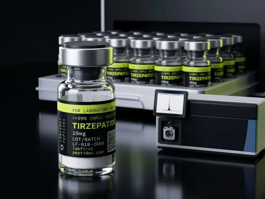
For research peptides the practical payoff of all this is a pair of numbers that beginners conflate. Chromatographic purity states what fraction of the detected material sits in the main HPLC peak. Net peptide content states what fraction of the vial's gross weight is peptide at all, as opposed to the counterions, usually trifluoroacetate or acetate, and the residual water that lyophilized material always carries. A vial labeled 10 mg at 99 percent purity may hold about 8 mg of actual peptide, and both figures can be honest at once because they answer different questions. Content is established by amino acid analysis or nitrogen determination. Purity never can be, because chromatography only sees what dissolves and absorbs.
Reading the two numbers in order keeps them apart.
- Read chromatographic purity: the fraction of detected material in the main HPLC peak.
- Read net peptide content: the fraction of the vial's gross weight that is peptide at all, as opposed to counterions and residual water.
- Check how content was established. Amino acid analysis or nitrogen determination can give it; chromatography never can, because it only sees what dissolves and absorbs.
- Calculate concentration from net peptide, not gross weight. A concentration calculated from gross vial weight overstates the true value by whatever the content discount is, and the error propagates silently into every figure derived from it.
The vial concentration calculator handles the mg-per-mL arithmetic for laboratory documentation; the documentation a supplier should stand behind, and how to read it, is set out in the quality standard. What happens to any of these molecules once water is added is a separate subject with its own rules, covered in the storage and stability guide.
FOR LABORATORY AND IN-VITRO RESEARCH USE ONLY. NOT FOR HUMAN OR ANIMAL CONSUMPTION. NOT FOR PERSONAL, MEDICAL, DIAGNOSTIC, THERAPEUTIC, OR RECREATIONAL USE.
Bottom line
An amino acid is one monomer of 75 to 204 Da; a protein is a folded chain of dozens to thousands of them; a peptide sits between, long enough to have a sequence and usually too short to hold a fold. Every peptide bond forms with the loss of one water, so a chain's mass is its residue masses plus 18 Da. Free amino acids are measured by amino acid analysis, peptides by HPLC paired with mass spectrometry, bulk protein by assays that count nitrogen or absorbance. On a certificate, purity and net peptide content answer different questions, and the arithmetic uses the second one.
Common questions
Is a peptide just a small protein?
How many amino acids exist compared with how many build proteins?
Why are sequences written from the N-terminus?
Why does a bulk protein assay give the wrong answer for a short peptide?
What is the difference between purity and peptide content?
Related guides: peptide chemistry basics
Sources
- IUPAC-IUBMB Joint Commission on Biochemical Nomenclature, amino acid and peptide nomenclature recommendations. Fixes the three-letter and one-letter residue codes, the residue concept, and the N-terminus-to-C-terminus writing convention used throughout.
- AOAC International official methods for nitrogen determination (Kjeldahl and Dumas). Basis of the nitrogen-times-6.25 protein estimate, its 16 percent nitrogen assumption, and the limits that follow from measuring nitrogen rather than protein.
- PubChem compound records for the proteinogenic amino acids. Monomer structures, formulas and masses quoted here, including glycine at 75 Da and tryptophan at 204 Da.
- Moore and Stein, classical ion-exchange amino acid analysis. Reference method behind composition analysis and net peptide content; the post-column ninhydrin arrangement descends from this work.